A recent study has shown the potential of tooth tissue-derived organoids have the potential to induce amelogenesis when exposed to the correct molecular signals and growth factors.
By Dr. Mehmood Asghar
Dental Tissues Have Limited Regenerative Potential
Unlike other body organs with relative degrees of regenerative potential, dental tissues, especially the dental enamel, have limited regenerative ability. Hence, despite vigorous efforts to regenerate damaged dental tissues and to overcome the need for dental restorations with limited clinical lifespans, these efforts have largely been successful in the laboratories – and their translation to the clinical scenario is still being investigated.
Although mesenchymal cells such as the dental pulp and periodontal ligament (PDL) have been extensively characterized, information regarding the presence, phenotype, and biological functions of epithelial stem cells (ESCs) are insufficient. However, previous research has shown that ESCs are present in the epithelial rest cells of Malassez (ERM) – an epithelial network of cells present in the dental follicle that encloses an unerupted tooth – and continues to remain in the PDL around the tooth root even after exfoliation (Davis, 2018).
Dental Epithelial Cells Could Potentially Regenerate Dental Enamel
Interestingly, these epithelial cells have been shown to possess some regenerative potential, owing to stem cell-related markers. These epithelial cells may then regenerate enamel by redirecting their growth to differentiate into ameloblasts – the epithelial cells that form the dental enamel. But unfortunately, the regenerative potential of these cells is significantly limited postnatally (Hamamoto et al., 1996).
Previous studies showed that the co-culturing of ERM-derived stem cells with dental pulp stem cells (DPSCs) could induce ameloblast differentiation. However, these 2D cultured cells showed limited growth potential and rapid loss of phenotype (Athanassiou-Papaefthymiou et al., 2015; Hyun et al., 2014). Hence, to grow dental tissues, the long-term phenotypical stability and growth potential of stem cells are desired.
The Organoid Technology
The problem of rapid phenotype and growth potential loss in dental stem cells could be overcome with organoid technology (Boretto et al., 2017; Schutgens & Clevers, 2020). Organoids are 3D constructs that can be self-developed when seeded in an extracellular matrix (ECM) – mimicking scaffold (Matrigel) and subsequently cultured in a pre-defined cocktail of growth factors (GFs), replicating cell signaling pathways or tissue embryogenesis. Under specific culture conditions, these organoids have been shown to duplicate the molecular and phenotypical characteristics of the stem cell compartment of the tissue of origin and could potentially be differentiated into desired tissue cells.
A significant benefit of the organoids is that they can be serially expanded (passaged) without loss of phenotype and regrowth potential, hence offering a powerful tool that overcomes the limited regenerative potential of the primary epithelial tissue stem cells.
Organoid Technology for Regeneration of Dental Tissues: The Groundbreaking Research
Although organoids have previously been derived for various body organs, epithelial stem cells for the dental tissues have not been established or characterized in detail yet. Recently, a group of scientists from KU Leuven and Hasselt University achieved a breakthrough in using dental epithelial cells to regenerate dental enamel. This research was published in the Cellular and Molecular Life Sciences Journal (Hemeryck et al., 2022).
The Methodology
This study was performed at the Oral and Maxillo-Facial Surgery – Imaging and Pathology (OMFS-IMPATH) unit of University Hospital Leuven. Predominantly unerupted third molars were extracted from adolescent patients.
The gingiva was separated to obtain the DF tissue, and the third molars – and their associated DFs – were isolated following bone perforation. Afterward, the DF tissue was peeled off the tooth and collected in Eagle’s Minimum Essential Medium (αMEM), containing 10% fetal bovine serum, 1% penicillin-streptomycin, and 0.5% fungizone. The tissue was then minced into small fragments and further dissociated using collagenase VI and Dispase II for 2 hours at 37 °C. Finally, a collection of single cells or small clusters was obtained using a 40 40 µm cell strainer.
The dissociated DF cells were resuspended in a mixture of serum-free defined medium (SFDM) and growth factor-reduced Matrigel in a 30:70 ratio and placed in 48-well plates (20,000 cells/ 20 µL drop). The tooth organoid medium (TOM) medium was supplemented where indicated upon solidification. Organoid cultures were kept at 37 °C in a 1.9% CO2 incubator, and the medium was refreshed every 2–3 days, each time supplemented with fungizone (0.1%). The organoid cultures were passaged every 10-14 days.
The Findings
1. DF-Derived Organoids Possess Regenerative Potential
The DF-derived organoids developed in this study showed a stemness profile similar to ERM. Interestingly, when the organoid cells were cultured, they were shown to induce the proliferation of ERM stem cells, highlighting the potential for tissue regeneration. Furthermore, single-cell transcriptomics – besides reinforcing the organoid-ERM congruence – uncovered novel molecular pathways previously observed in mice.
2. Organoid-Induced Stem Cell Differentiation Follow In-vivo Pathways
When the organoids were exposed to the epidermal growth factor (EGF), it led to the transient proliferation and subsequent epithelial-mesenchymal transition. These changes highly mimic the events that take place in ERM in-vivo.
3. TGFβ Induces Amelogenesis Differentiation in Organoids
The ERM stemness organoids were able to initiate an ameloblast differentiation process further enhanced by the transforming growth factor β (TGFβ) and abrogated upon TGFβ receptor inhibition. This also reconfirms the critical role of TGFβ in amelogenesis. In addition, potent signs of remineralization were observed in MIM- compared to TOM-cultured organoids – as evidenced through transmission electron microscopy. Interestingly, the remineralization was increased in the organoids when cultured in-vivo following subcutaneous transplantation in 3D-printed hydroxyapatite scaffolds in immunodeficient mice.

Clinical significance
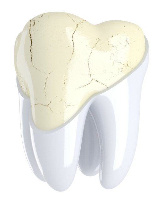
Dental enamel is the hardest tissue of the body, and it plays a vital role in keeping our teeth sensitivity- and cavity-free. Unfortunately, it does not have regenerative potential. Once lost, the only restorative option is topical remineralization – in case of partial loss – or dental fillings when completely damaged. However, these options have varying levels of efficacy.
The findings of this study have opened a pathway whereby dental organoids could be used to induce stem cell regeneration into various dental tissues. Besides, these organoids also have the potential to decipher the molecular pathways in tooth development and, more importantly, dental pathogenesis. Hence, dental organoid technology can be used to treat various dental infections and genetic anomalies like amelogenesis imperfecta.
Future research needs to emphasize translating these findings from laboratories toward clinical trials, paving the way for the realization of dental tissue regeneration and lesser reliance on dental restorations and prosthetics.
Author: Dr. Mehmood Asghar is a dentist by profession and an Assistant Professor of Dental Biomaterials at the National University of Medical Sciences, Pakistan. Dr. Asghar received his undergraduate and postgraduate dental qualifications from the National University of Science and Technology (NUST). He is also currently pursuing a Ph.D. in Restorative Dentistry from Malaysia. Apart from his hectic clinical and research activities, Dr. Asghar likes to write evidence-based, informative articles for dental professionals and patients. Dr. Asghar has published several articles in international, peer-reviewed journals.
References
Athanassiou-Papaefthymiou, M., Papagerakis, P., & Papagerakis, S. (2015). Isolation and characterization of human adult epithelial stem cells from the periodontal ligament. Journal of Dental Research, 94(11), 1591-1600.
Boretto, M., Cox, B., Noben, M., Hendriks, N., Fassbender, A., Roose, H., Amant, F., Timmerman, D., Tomassetti, C., Vanhie, A., Meuleman, C., Ferrante, M., & Vankelecom, H. (2017). Development of organoids from mouse and human endometrium showing endometrial epithelium physiology and long-term expandability. Development, 144(10), 1775-1786.
Davis, E. M. (2018). A review of the epithelial cell rests of malassez on the bicentennial of their description. Journal of Veterinary Dentistry, 35(4), 290-298.
Hamamoto, Y., Nakajima, T., Ozawa, H., & Uchida, T. (1996). Production of amelogenin by enamel epithelium of hertwig's root sheath. Oral Surgery, Oral Medicine, Oral Pathology, Oral Radiology and Endodontics, 81(6), 703-709.
Hemeryck, L., Hermans, F., Chappell, J., Kobayashi, H., Lambrechts, D., Lambrichts, I., Bronckaers, A., & Vankelecom, H. (2022). Organoids from human tooth showing epithelial stemness phenotype and differentiation potential. Cellular and Molecular Life Sciences, 79(3), 153.
Hyun, N., Ji-Hye, K., Jae-Won, K., Byoung-Moo, S., Joo-Cheol, P., Jung-Wook, K., & and Gene, L. (2014). Establishment of hertwig’s epithelial root sheath/epithelial rests of malassez cell line from human periodontium. Molecules and Cells, 37(7), 562-567.
Schutgens, F., & Clevers, H. (2020). Human organoids: Tools for understanding biology and treating diseases. Annual Review of Pathology: Mechanisms of Disease, 15(1), 211-234.